Anatomy Rib Cage Muscles - Pin on Twisted yoga. Rib cage joint rib cage stretches rib cage art rib cage abnormalities rib cage workouts rib cage and abdominals rib cage components rib cage muscle tear rib cage injury treatment. All muscles that are attached to the human rib cage have the. The muscles that affect the knee's movement run along the thigh and calf. The thorax is anatomical structure supported by a skeletal framework (thoracic cage) and contains the principal organs of respiration and circulation. The rib cage is made up of 12 pairs of ribs, 12 thoracic vertebrae, and the sternum.
Rib cage anatomy and breathing. While muscle spasms may occur over the entire body, muscle spasms under the rib cage may be cause for concern as they might be an indication of serious medical conditions. They are attached to the femur (thighbone), tibia (shinbone), and fibula (calf bone) by fibrous tissues called ligaments. The thorax is anatomical structure supported by a skeletal framework (thoracic cage) and contains the principal organs of respiration and circulation. They are each attached to.
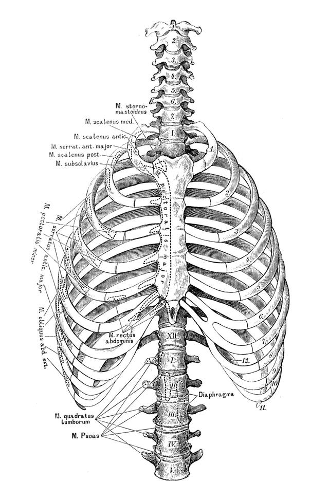
Human Muscle Body With Ribs Stock Illustration - Image ... from thumbs.dreamstime.com How do you build muscles on your rib cage? Seventeen muscles attach to the scapula, and it articulates with the clavicle to form the shoulder girdle or pectoral girdle, which supports movements. During normal breathing, contraction of the major inspiratory muscle, the diaphragm, produces both rib cage expansion and a downward movement of the diaphragm. Contributing to their role in protecting the internal thoracic organs. Learn anatomy faster and remember everything you learn. The muscles that affect the knee's movement run along the thigh and calf. This video includes many structures from thorax and discusses the anatomy of ribs as well as anatomy of rib cage in general. The ribcage is made to be flexible and springy so the lungs can fill and deflate easily.
They are more involved in forced expiration and coughing to forcibly shrink the chest and.
Create your own flashcards or choose from millions created by other students. See more ideas about anatomy, anatomy study, rib cage anatomy. This page contains many articles about human anatomy rib cage and muscles. Each rib articulates posteriorly with the vertebral column. Learn anatomy faster and remember everything you learn. They can move the ribs and have roles in breathing. Structure of a typical rib: The rib cage is a primarily protective structure, encircling the heart and lungs. In the back, latissimus dorsi and erector spinae muscles (anatomy lesson #10) cover the 11th and 12th ribs of the thoracic cage and deeper yet are the paired abdominal kidneys flanking the. Rib cage anatomy in homo erectus suggests a recent evolutionary origin of modern human body shape. The primary responsibilities of the ribcage involve protecting the thoracic visceral organs, enclosing the thoracic visceral organs, and is included in the general mechanics of the process of breathing. The thorax resembles a truncated cone which is. Everyone has nice muscles in ct scanning!
They can move the ribs and have roles in breathing. Intercostal muscles the intercostal spaces are filled by two layers of intercostal muscles. A great aid is a medical anatomy atlas ( the kind that is mostly pictures, color is. The muscles that affect the knee's movement run along the thigh and calf. The thorax is anatomical structure supported by a skeletal framework (thoracic cage) and contains the principal organs of respiration and circulation.
How Many Ribs do Humans Have? from pixfeeds.com Top suggestions for rib cage anatomy muscles. The human rib cage (thoracic cage) has the very important job of protecting the heart and lungs. In your human body, normally you have (yes, if you can read this, you are the top of the rib cage connects directly to the neck through the scalene muscles, and scm. Quizlet is the easiest way to study, practise and master what you're learning. The muscles that affect the knee's movement run along the thigh and calf. Structure of a typical rib: This video includes many structures from thorax and discusses the anatomy of ribs as well as anatomy of rib cage in general. During normal breathing, contraction of the major inspiratory muscle, the diaphragm, produces both rib cage expansion and a downward movement of the diaphragm.
The rib cage is a primarily protective structure, encircling the heart and lungs.
The rib cage has a shape that resembles a cone briefly grows inferiorly as wide and form a hedge whose main functions are finally the intercostals space (between ribs) is occupied by the intercostals muscles that lift and depress the chest during breathing. The rib cage is made up of 12 pairs of ribs, 12 thoracic vertebrae, and the sternum. Rib cage muscles and tendons / spinal anatomy and back pain : Everyone has nice muscles in ct scanning! The intercostal muscles extend from the. Similarly, if a person has experienced trauma to the muscles or ligaments in the chest, there is not much that can be done to reduce movement—as the chest needs to move at least enough to expand. Beginning with one of the most dominant muscles in the region, the trapez. Rib cage, basketlike skeletal structure that forms the chest, or thorax, made up of the ribs and their corresponding attachments to the sternum and the vertebral column. The primary responsibilities of the ribcage involve protecting the thoracic visceral organs, enclosing the thoracic visceral organs, and is included in the general mechanics of the process of breathing. It provides a strong framework onto which the muscles of the cramps in ribcage are often observed in those who strain or overwork their upper body. Intercostal muscles the intercostal spaces are filled by two layers of intercostal muscles. They are attached to the femur (thighbone), tibia (shinbone), and fibula (calf bone) by fibrous tissues called ligaments. Learn anatomy faster and remember everything you learn.
Struggling with learning muscle attachments? Similarly, if a person has experienced trauma to the muscles or ligaments in the chest, there is not much that can be done to reduce movement—as the chest needs to move at least enough to expand. Another important feature of the rib cage is the manubriosternal joint also known as the sternal angle of louis. It provides a strong framework onto which the muscles of the cramps in ribcage are often observed in those who strain or overwork their upper body. Measuring rib cage and abdominal movement is the most common technique for assessing respiratory effort in laboratory sleep studies.
anatomy IV at University of New England - StudyBlue from classconnection.s3.amazonaws.com The human rib cage (thoracic cage) has the very important job of protecting the heart and lungs. Contributing to their role in protecting the internal thoracic organs. The primary responsibilities of the ribcage involve protecting the thoracic visceral organs, enclosing the thoracic visceral organs, and is included in the general mechanics of the process of breathing. • raise rib cage for inhaling & depresses rib cage for exhaling. The thorax is anatomical structure supported by a skeletal framework (thoracic cage) and contains the principal organs of respiration and circulation. Rib cage anatomy in homo erectus suggests a recent evolutionary origin of modern human body shape. Tendons attach the muscles to each other. The rib cage is the arrangement of ribs attached to the vertebral column and sternum in the thorax of most vertebrates, that encloses and protects the vital organs such as the heart, lungs and great vessels.
Similarly, if a person has experienced trauma to the muscles or ligaments in the chest, there is not much that can be done to reduce movement—as the chest needs to move at least enough to expand.
Intercostal muscles internal and external view. Create your own flashcards or choose from millions created by other students. Rib cage muscles and tendons / spinal anatomy and back pain : The ribs are a set of twelve paired bones which form the protective 'cage' of the thorax. The intercostal muscles lie between the ribs and build the walls of the thorax. Struggling with learning muscle attachments? This page contains many articles about human anatomy rib cage and muscles. Rib cage anatomy and breathing. • raise rib cage for inhaling & depresses rib cage for exhaling. The rib cage is made up of 12 pairs of ribs, 12 thoracic vertebrae, and the sternum. The ribcage is made to be flexible and springy so the lungs can fill and deflate easily. The muscles that affect the knee's movement run along the thigh and calf. Learn anatomy faster and remember everything you learn.
The thorax resembles a truncated cone which is anatomy rib cage. A rib has a flat body.
Share this post
0 Response to "Anatomy Rib Cage Muscles - Pin on Twisted yoga"


0 Response to "Anatomy Rib Cage Muscles - Pin on Twisted yoga"
Post a Comment